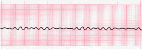
Ventricular Fibrillation ECG Interpretation #315
Description
- The morphologic features are different with this dysrhythmia. No P wave and no QRS complexes. This rhythm presents with a chaotic waveform which reflects the electrical chaos occurring within the heart.
- The heart is not actually beating as we know it. The chaos occurs as a result of small regions of tissue which are independently depolarizing.
- This rapid disorganized electrical activity actually makes the heart appear to quiver in response to this activity. Some have described it as shaking like Jello.
- Fibrillatory waves may be coarse or very fine. This is based upon their size. The longer V Fib occurs, the smaller the waveforms are likely to be.
- (Description continues on next slide)

Description (continued)
- Coarse Ventricular Fibrillation (coarse V Fib) is when a majority of the waveforms measure 3 mm or greater
- Fine V Fib (fine vfib) is when a majority of the waveforms measure less than 3 mm
- This is absolutely a life-threatening dysrhythmia which requires, immediate, effective, and aggressive care.
- (Follow your local reporting and treatment protocols)
- If your patient is talking to you when you see this on the monitor, then your patient is not in V Fib. Always, check your patient first, but there will likely be a loose or disconnected lead wire or electrode.

VFib Practice Strip

Analyze this vfib rhythm strip using the five steps of rhythm analysis.
Show Answer
- Rhythm: Irregular
- Rate: Unable to determine
- P Wave: absent
- PR interval: absent
- QRS: absent
- Interpretation: Ventricular Fibrillation (Fine)
Authors and Sources
Authors and Reviewers
- ECG heart rhythm modules: Thomas O'Brien.
- ECG monitor simulation developer: Steve Collmann
-
12 Lead Course: Dr. Michael Mazzini, MD.
- Spanish language ECG: Breena R. Taira, MD, MPH
- Medical review: Dr. Jonathan Keroes, MD
- Medical review: Dr. Pedro Azevedo, MD, Cardiology
- Last Update: 11/8/2021
Sources
-
Electrocardiography for Healthcare Professionals, 6th Edition
Kathryn Booth and Thomas O'Brien
ISBN10: 1265013470, ISBN13: 9781265013479
McGraw Hill, 2023 -
Rapid Interpretation of EKG's, Sixth Edition
Dale Dublin
Cover Publishing Company -
EKG Reference Guide
EKG.Academy -
12 Lead EKG for Nurses: Simple Steps to Interpret Rhythms, Arrhythmias, Blocks, Hypertrophy, Infarcts, & Cardiac Drugs
Aaron Reed
Create Space Independent Publishing -
Heart Sounds and Murmurs: A Practical Guide with Audio CD-ROM 3rd Edition
Elsevier-Health Sciences Division
Barbara A. Erickson, PhD, RN, CCRN -
The Virtual Cardiac Patient: A Multimedia Guide to Heart Sounds, Murmurs, EKG
Jonathan Keroes, David Lieberman
Publisher: Lippincott Williams & Wilkin)
ISBN-10: 0781784425; ISBN-13: 978-0781784429 - Project Semilla, UCLA Emergency Medicine, EKG Training Breena R. Taira, MD, MPH
-
ECG Reference Guide
PracticalClinicalSkills.com