This is an example of moderate mitral stenosis. The first heart sound is increased in intensity. The second heart sound is normal and unsplit. Systole is silent. There is an opening snap 75 milliseconds into diastole. As mitral stenosis becomes more severe, this opening snap will occur earlier in diastole.
A diamond-shaped, low-frequency murmur follows the opening snap. There is a second murmur in late diastole due to contraction of the left atrium.
Auscultate using the bell of the stethoscope.
Mitral stenosis can be caused by rheumatic heart disease, and this causes a thickening of the mitral valve leaflets. In the cardiac animation, observe turbulent blood flow from the left atrium into the left ventricle. Take note of the moderately thickened mitral valve leaflets and the moderately enlarged left atrium. The excursion of the mitral valve leaflets is moderately decreased.
Listen
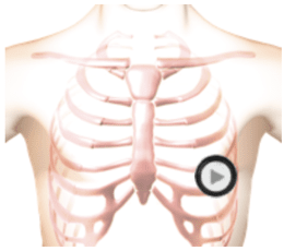

The patient's position is supine left side down.