This lesson presents severe mitral regurgitation. The first heart sound is normal while the second heart sound is widely split. A third heart sound gallop follows S2. A loud, rectangular, pansystolic murmur is present. In addition, a brief, rumbling, diamond-shaped diastolic murmur immediately after the third heart sound.
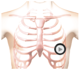
In the cardiac anatomy video, you can see the enlarged left atrium and left ventricle. Observe the turbulent blood flow from the left ventricle into the left atrium. This is the systolic murmur. Also notice the brief turbulent blood flow from the left atrium to the left ventricle during diastole. This is caused by too much blood in the left atrium which forces blood back into the ventricle during diastole producing the flow rumble. Severe mitral regurgitation is often caused by degeneration of the mitral valve leaflets.
Listen
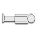
The patient's position is supine.