Atrial Septal Defect is a congenital condition associated with abnormal blood flow between the left atrium and the right atrium. Before birth there is a large connection between right and left atria. During fetal development, the connection gradually disappears. However, in some cases, this opening persists and is known as an atrial septal defect.
Both the first and second heart sounds are split. The second heart sound splitting is fixed at 80 milliseconds. There is a brief diamond-shaped murmur in early systole and another brief diamond-shaped murmur in early diastole
This murmur was auscultated at the pulmonic position.
In the cardiac animation video, observe an enlarged right atrium and right ventricle. You see turbulent blood flow across the tricuspid valve between the right atrium and the right ventricle (the diastolic murmur). This is caused by blood flow from the left atrium into the right atrium through the atrial septal defect. There is additional turbulent flow into the pulmonary artery causing the systolic murmur.
Listen
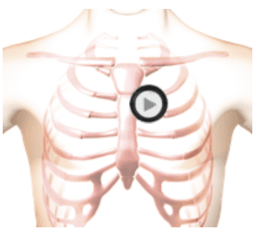
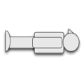
The patient's position is supine.