ECG Axis Determination Module
ECG Axis Determination
Thomas E. O'Brien
AS CCT CRAT RMA
Learning Objectives
Upon completion of the accompanying narrative and practice the student will be able to:
- Recall which major coronary artery supports each region of the heart
- Associate the views of a 12-Lead ECG with specific surfaces of the heart
- Determine the presence of cardiac axis deviation
- Understand Einthoven's Triangle
12-Lead ECG
- People may choose to analyze ECG’s in a number of different ways. The sequence doesn’t necessarily matter as long as you gather and report the proper information each time. I read from left to right as much as possible (in anatomic groupings).
- If your protocols are different, always follow them.
- The following slides will review the leads, surfaces, and associated coronary arteries which commonly supply that portion of the heart.
Please sign in. This module is premium content.
Lessons
Lesson #1: Views 322
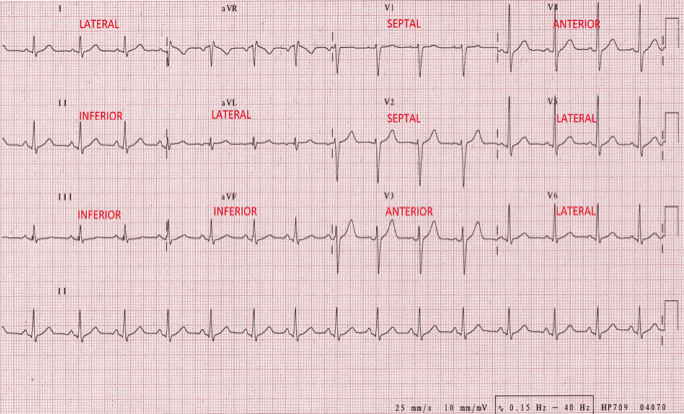
II, III, and aVF
- Inferior wall of left ventricle
- Right Coronary Artery
- Marginal branch
V1 and V2
- Septum
- Left Coronary
- Septal branch
V3 & V4
- Anterior
- Left Coronary
- Anterior Descending and Diagonal arteries
I, aVL, V5 & V6
- Lateral
- Left Coronary
- Circumflex & Obtuse marginal
Labeled Views
Lesson #2: Quick Check Views 1
Question 1
Name the inferior view leads.
Answer
II, III & aVF
Question 2
Name the septal view leads.
Answer
V1 & V2
Lesson #3: Quick Check Views 2
Question 1
Name the anterior view leads.
Answer
V3 & V4
Question 2
Name the lateral view leads.
Answer
I, aVL, V5 & V6
Lesson #4: Axis & Deviation
Description
- Axis is the general flow of electricity as it passes through the heart from the SA node all the way to the Purkinje fibers.
- It is typically obtained from the limb or frontal plane leads.
- The axis of the heart can change for a number of different reasons from birth defects which affect the physical location of the heart i.e. Dextrocardia, to obesity to pregnancy among others.
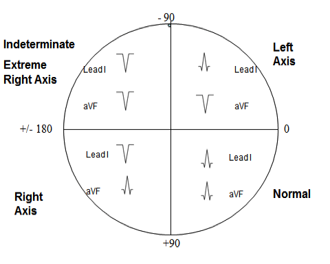
- If you can envision a circle with a centered vertical and horizontal line superimposed over the chest, you would note that the normal heart is located in the lower left quadrant of this circle.
- You will be provided an example of this image in a few slides.
Electrical Axis Determination
Axis determination can be determined a number of different ways. From complex to simple. The following steps are the easiest I know to obtain this important information.
Electrical Axis Determination
Imagine a large circle, divided into four equal quadrants superimposed over the patient’s chest.
Lesson #5: Axis Determination
Part I
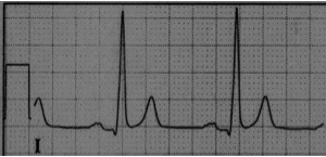
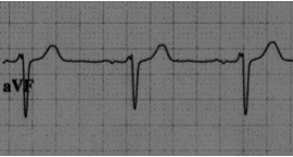
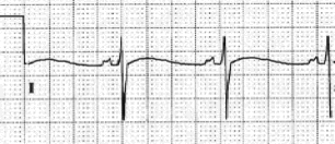
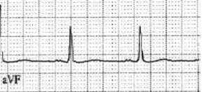
- The direction of the ventricular depolarization (QRS complex) is analyzed.
- Refer to lead I and aVF each time
- If the majority of the QRS complex is positively deflected in both views, this is referred to as “normal axis”.
Part II
If lead I is positive and aVF is negative, this is ‘left axis deviation”
Part III
If lead I is negative and aVF is positive, this is “right axis deviation”.
Part IV
If lead I is negative and aVF is negative, this is extreme right axis deviation aka indeterminate.
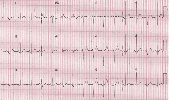
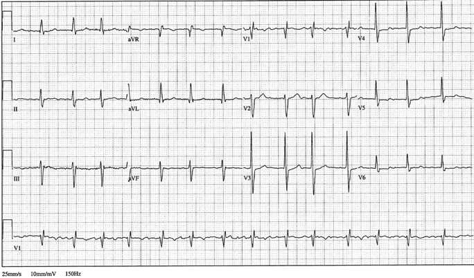
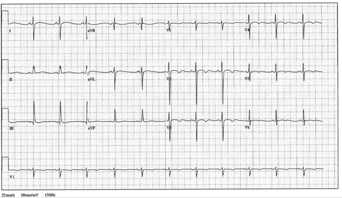
Lesson #6: Quick Check Axis 1
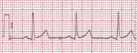
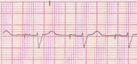
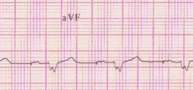
Question
Identify the electrical axis
Answer
Normal Axis – Lead I up, aVF up
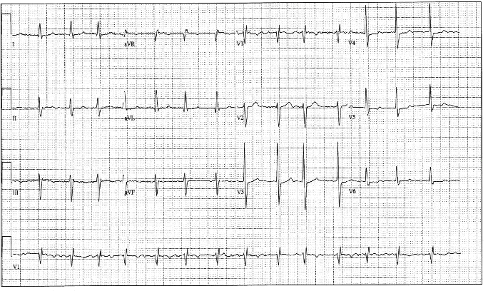
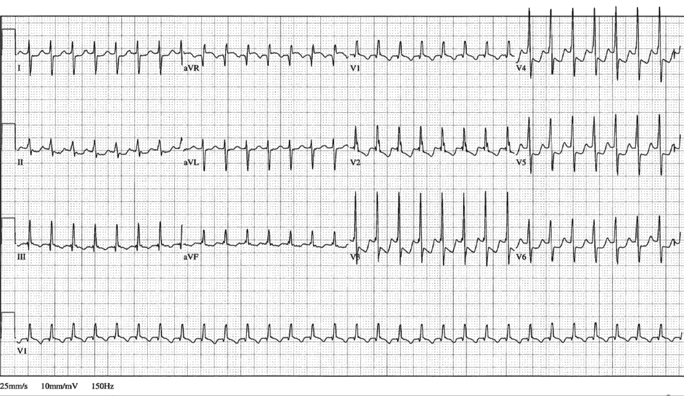
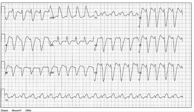
Question
Identify the electrical axis
Answer
Despite the presence of premature ventricular complexes, when analyzing the ventricular depolarization of the underlying rhythm you will note Normal Axis – Lead I up, aVF up
Question
Identify the electrical axis
Answer
Normal Axis – Lead I up, aVF up
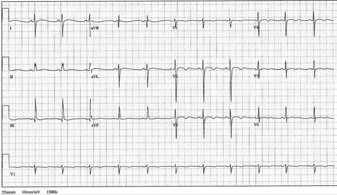
Lesson #7: Quick Check Axis 2
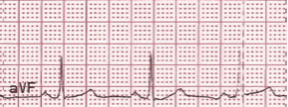
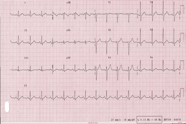
Question
Identify the electrical axis
Answer
Left Axis – Lead I up, aVF down
Question
Identify the electrical axis
Answer
Left Axis – Lead I up, aVF down
Question
Identify the electrical axis
Answer
Left Axis – Lead I up, aVF down
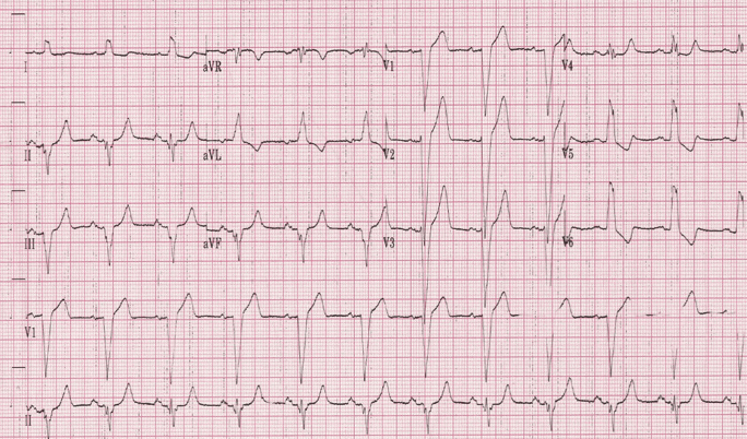
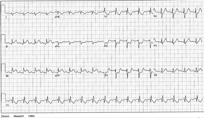
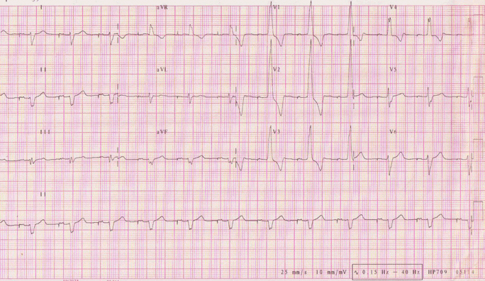
Lesson #8: Quick Check Axis 3
Question
Identify the electrical axis
Answer
Left Axis – Lead I up, aVF down
Question
Identify the electrical axis
Answer
Right Axis – Lead I down, aVF up
Question
Identify the electrical axis
Answer
Right Axis – Lead I down, aVF up
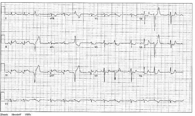
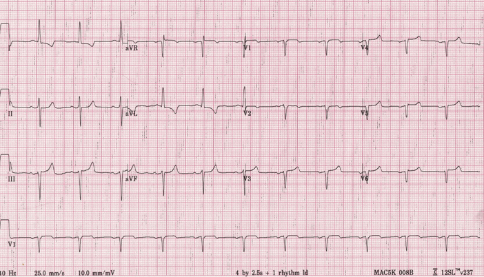
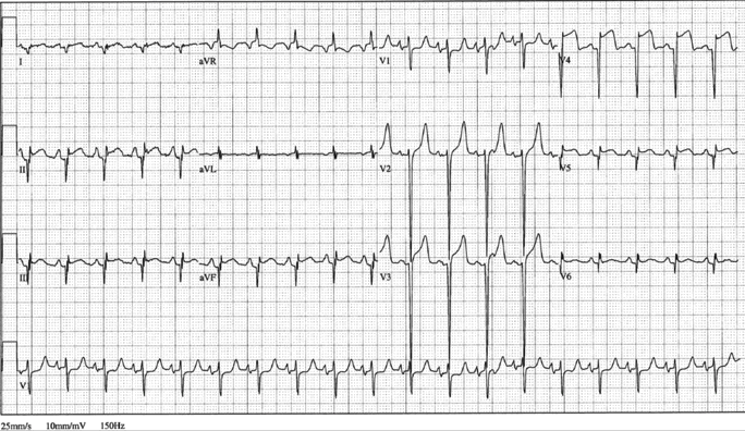
Lesson #9: Quick Check Axis 4
Question
Identify the electrical axis
Answer
Right Axis – Lead I down, aVF up
Question
Identify the electrical axis
Answer
Right Axis – Lead I down, aVF up
Question
Identify the electrical axis
Answer
Extreme Right Axis – Lead I down, aVF down
Lesson #10: Quick Check Axis 5
Question
Identify the electrical axis
Answer
Extreme Right Axis – Lead I down, aVF down
Question
Identify the electrical axis
Answer
Right Axis – Lead I down, aVF up
Authors and Reviewers
- EKG heart rhythm modules: Thomas O'Brien
-
Medical review: Dr. Jonathan Keroes, MD
- Medical review: Dr. Pedro Azevedo, MD, Cardiology
-
Last Update: 11/8/2021
Sources
-
Electrocardiography for Healthcare Professionals, 6th Edition
Kathryn Booth and Thomas O'Brien
ISBN10: 1265013470, ISBN13: 9781265013479
McGraw Hill, 2023 -
Rapid Interpretation of EKG's, Sixth Edition
Dale Dublin
Cover Publishing Company -
EKG Reference Guide
EKG.Academy -
12 Lead EKG for Nurses: Simple Steps to Interpret Rhythms, Arrhythmias, Blocks, Hypertrophy, Infarcts, & Cardiac Drugs
Aaron Reed
Create Space Independent Publishing -
The Virtual Cardiac Patient: A Multimedia Guide to Heart Sounds, Murmurs, EKG
Jonathan Keroes, David Lieberman
Publisher: Lippincott Williams & Wilkin)
ISBN-10: 0781784425; ISBN-13: 978-0781784429 -
ECG Reference Guide
PracticalClinicalSkills.com