Pelvis
Pelvis - Transverse

Narration
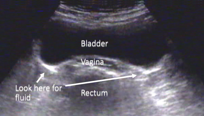
So in addition to the left and right upper quadrants, we can look into the pelvis to see peritoneal fluid. Typically we will start with a transverse view, with the indicator towards the patient’s right and right over the symphysis pubis scanning down and you will see fluid here, but it is the bladder so there’s a full bladder of black fluid and you’ll notice its encapsulated and not tracking into those crevasses that we talk about with free fluid.
Pelvis - Transverse


Narration
This is a diagram showing the bladder, where you would look for fluid, the vagina, the rectum, and again this is a transverse scan with the indicator to the patient’s right and fluid in the bladder, but no free fluid.
Pelvis – Sagittal

Narration
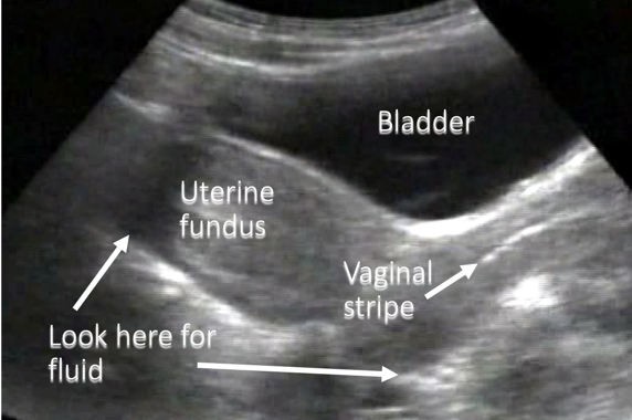
We can also then rotate the transducer so the indicator is towards the head and in this case, we’re interrogating the female pelvis with the uterus posterior to the bladder and this is normal.
Pelvis – Sagittal


Narration
We’re looking at the vaginal stripe which is the potential space between the anterior and posterior vaginal walls going up to the cervix, the uterine fundus, the bladder which is anterior, and then we are looking for fluid typically behind the uterus either in the pouch of Douglas which is more inferior or to the right of the screen, or towards the top, over the top of the uterus.