Other Fluid & Summing Up
Fluid - Hydrocele
Narration
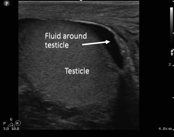
So once we understand the principles of finding fluid, we can use it in other places. This is an example of a scan of the testicle actually. We are using a linear probe - it’s quite easy, there’s no bones or anything else in the way, we can see the regular testicular architecture along with the black fluid surrounding it.
Fluid - Hydrocele

Narration
That is known as a hydrocele, just fluid around the testicle.
Contained Fluid - Cyst

Narration
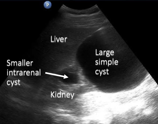
We can also find fluid that is encapsulated within a structure such as a cyst. This is an example of two simple cysts, one that’s very large and on the inferior pole of the kidney. This one’s probably about 8cm, and there’s also a small simple cyst within the kidney.


Narration
Again, a simple cyst is completely anechoic, thin walled, and doesn’t have any debris or septations within it.

Narration
We may see more complex cystic structures that do have fluid within them. In this case this is actually a hepatic cyst that ruptured and you can see debris, maybe blood, septations along with the fluid, and this is a complex cyst.
Fluid - Abscess
Narration
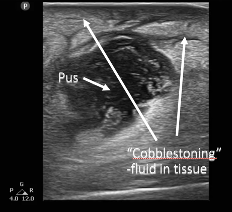
Other places we may see fluid include soft tissue and this is an examination of an abscess. You can see that there is a black area of fluid, in this case pus.
Fluid - Abscess

Narration
It’s completely anechoic – it’s got some debris in it and you can also see some fluid within the interstitium of the fatty tissue and this is called cobblestoning or fluid within the tissue.
Summing Up – Finding Fluid
- Track fluid
- Which space?
- Contained or free?
- Simple or complex?
Narration
So to sum up, when you find black, it looks to be fluid you track it and see where it goes. You look at this picture on the right it’s a Morison’s pouch view, and there’s actually fluid both in Morison’s pouch between the liver and the kidney, as well as in the thoracic space above the diaphragm. The key to finding fluid and knowing where it is and what significance it has is to track it to see if its contained or free, to see if its simple or complex, and basically make sure you know exactly what it entails, where it is, and how much of it you may see.