The Inferior Thoracic Space
Inferior Thoracic Space For Effusion
- Coronal plane
- Slide/ angle superiorly
- Visualize diaphragm
- Look for fluid above

Narration
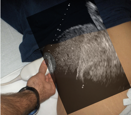
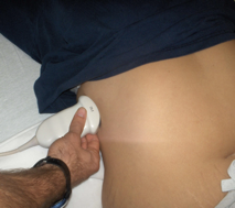
So we’ll start with the inferior thoracic spaces. In this case we’re using a curvilinear probe at the right flank. This is a coronal plane and we’re looking into the body. Typically you will see the liver here and above the liver is the diaphragm which is where you are really looking to interrogate. You just angle the probe a little bit more superiorly than you would for the liver and kidney and try to get that inferior thoracic space.
Inferior Thoracic Space For Effusion
- Coronal plane
- Slide/ angle superiorly
- Visualize diaphragm
- Look for fluid above

Narration
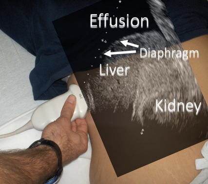
So when we do this, we’ll again see the liver, and above the liver is that bright or hyperechoic diaphragm and in this case we do see black above that, which is fluid or a pleural effusion.
Inferior Thoracic Space - Normal

Narration
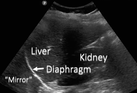
Now here is a moving image of a normal thoracic space. In this case we see the liver, the kidney, a couple of rib shadows, and we can see that diaphragm and actually on the other side of the diaphragm it almost looks like there’s liver, and this is what is called as a mirror image artifact – it’s actually normal and it tells you there is not an effusion or fluid above the diaphragm. It’s an artifact that is actually a reflection off the diaphragm and it actually tricks you into thinking it looks like the liver on the other side.
Inferior Thoracic Space - Normal


Narration
This still image shows that mirror image artifact again on the other side of the diaphragm - you really don’t see that black area that you might expect if there were fluid or a pleural effusion so were seeing the liver, the kidney, a rib shadow, the diaphragm, and mirror image artifact, which is normal.