Pleural Effusions
Phased Array

Narration
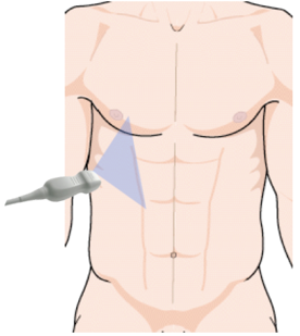
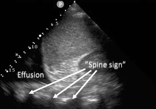
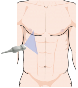
So here’s another example of interrogating the right inferior thoracic space in a coronal plane, in this case using a phased array probe placed at the right flank. You can tell it’s a phased array probe because it comes from that point at the top of the screen and this is actually a pretty helpful probe for getting in between ribs. In this case we can see the liver, kidney, the diaphragm, and you can see that triangular wedge shaped area of black which is fluid or pleural effusion and you can actually see what we call the spine sign, which is the proximal thoracic ribs or lateral vertebral processes extending up from behind the liver into the thoracic area.
Spine Sign


Narration
Here’s a labeled still image that still shows that spine sign. Its normal to see the spine behind the liver or the kidney, but if you continue to see it up behind the thoracic space in back of the effusion its actually the effusion that allows you to see that and helps you to know that there is an effusion, so this is what is called the spine sign.
Large Pleural Effusion

Narration
Here’s an example of a large pleural effusion on the right side. The diaphragm is very well delineated and you can see fluid above it and some consolidated or atelectatic lung kind of waving around in that pleural effusion.